|
The Microscope Lamp.
Design Considerations for the Ideal Köhler Illuminator. |
Page 5 of 5 |

|
The Microscope Lamp.
Design Considerations for the Ideal Köhler Illuminator. |
Page 5 of 5 |
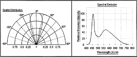
Since their development in the early nineties, white-light LEDs have made great progress. They are inherently more efficient than tungsten filament lamps in terms of the amount of electrical energy which they are able to convert into light. Whereas a tungsten lamp produces light at a rate of 20-25 Lumens per Watt, presently available white LEDs operate at 50 Lumens/Watt, and developers believe that with improvements of the phosphors used in their construction, this figure may be doubled again in the near future. Over the last three or four years, the light output of commercially available white LEDs has gone from 1000 to 7-8000 millicandelas. Whilst improvements in output are certain to continue, currently available white LEDs are more than equal to the task of replacing tungsten filament lamps in many, even most, light microscope applications. In the LED types considered here, the diode die itself produces blue light at a wavelength of about 470nm, and this excites a phosphor which produces yellow. The mixture of these two colours gives a fair approximation to a daylight which is unavoidably deficient in red. In spite of this, colour rendition with digital cameras is surprisingly good (see tests below). Power consumption is very low, and very little heat is generated. At an operating voltage of 3.6 V, the current is 30mA -- a total power rating of around 120mW, producing a light output of about 6000 mcd. 
From data sheet for narrow-beam white LED {below, left). Another attractive feature of these devices is their 50,000+ hours life expectancy. Whilst there is some fall-off of light output over time with the currently available LEDs due mainly to yellowing of the epoxy resin used in the package, this is not noticeable inside thousands of hours of usage. Experimenters should obtain a full specification for any LED intended for microscope use, as there are some varieties which generate a blue in the near ultraviolet at 380nm and could be harmful to the eyes when viewed directly as in a brightfield microscope, even allowing for the UV absorption of the microscope optics.
The pictures above (taken at the same magnification) show two similar-looking 5mm white LEDs, one of which is close to ideal as a microscope illuminant, and the other is all but unusable. The LED on the left has a small emitting area which is situated sufficiently far from the strongly curved lens moulded into the clear epoxy package for most of the emitted light to be concentrated into a beam of about 30°. When such an LED is placed at the principle focus of a wide aperture condenser lens having an angular aperture of about 90°, only the central portion of the lens is filled with light. Seen from the microscope, a central circle of about half the diameter of the condenser lens is illuminated. The view of the back lens of the objective is equally unsatisfactory, and it is near to impossible to fill both the lamp condenser and the aperture of a high-power objective simultaneously. True Köher illumination is therefore not achievable. 
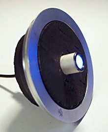
By contrast, the other LED (right, above) has a larger source placed close to the centre of curvature of its lens, producing a beam of remarkably even distrubution over an angle of 90° -- just large enough to fill a lamp condenser of 0.7 NA. Since the microscope field is filled with a highly enlarged portion of the emitting luminous source, and this source has an uneven distribution of blue and yellow light (in all examples of this LED examined), as well as images of the fine wires connecting the LED die to the external pins, some unevenness of colour and intensity is visible at the specimen plane. It can be reduced but not entirely eliminated by the introduction of some diffusion immediately in contact with the LED. This has been done in the assembly above, where the LED is a sliding fit in a metal sleeve which has a plastic film diffuser at its end. The following pictures have been shot using a Kodak DC4800 digital camera, the same optics (a x20 achromatic objective with Abbe condenser in a Köhler brightfield) and specimen (a stained cross-section of a Yew stem), but using LED (top pics) and tungsten filament (bottom pics) as light source. In both cases, the pictures were taken at a light level for comfortable viewing, giving shutter speeds of around 1/20th. sec. at ISO 100. The two leftmost pictures are with the camera's white-balance setting on "tungsten" (equivalent to 3200°K), and the two on the right were taken at the colour temperature setting on the camera which gave the most neutral/white backround. In pictures 3 and 4, the tungsten source was an Olympus/Hosobuchi 6V, 30W grid filament lamp with some blue filtration and an operating voltage of 3V to give a comfortable light level for visual use. This explains why the colour cast of picture 3 is so warm, even on the tungsten setting. No colour manipulations have been applied to any picture.
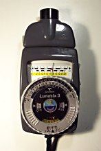
It can be seen that the picture with the cleanest white background and the most accurate colour rendition is given by the combination of LED illumination and camera set to 9000°K. The falloff in brightness in the lower right of the picture (more obvious in the picture than in visual use) is due to uneven blue/yellow distribution at the source. The accuracy of the green colour, as well as saturation in the reds is better than those of the tungsten illuminated picture. This is a surprising result given the spectral distribution of this LED. Nichia, the first company to make a white LED, has recently produced a The x20 brightfield setup described above was used to determine the brightness of the white LED relative to that of the Olympus 30W/6V tungsten filament lamp. The meter used was a Gossen Lunasix 3 with the microscope attatchment illustrated on the right.  The microscope eyepiece was replaced with the meter, and each illuminant was used in turn (with necessary lamp refocus adjustments) and an intensity reading made. The results showed that the filament lamp (at 6V) provided an illumination 4.5 photographic stops more intense than the LED (at 3.6V), or 16 - 32 times brighter.
Also of interest is the relative brightness of the white LED in a Köhler lamp, and a 60 Watt mains frosted light bulb sometimes used (in the absence of anything better) to light a laboratory microscope. Measurement showed that the LED in the Köhler lamp was 3.5 photographic stops brighter than the bulb. With the 60W bulb, a x40 power darkfield using an Abbe condenser with a substage stop is just bright enough for good observation. Since high-power darkfields of good saturation are probably the most light-demanding of microscope systems, it would seem that LEDs as microscope illuminants have well and truly arrived, but do not as yet offer as bright illumination as tungsten filament lamps designed for the job. With a few tweaks to the packaging of these devices, an LED ideally suited to microscopes could easily be produced. Within the constraints of a 5mm diameter package, the LED pictured above on the right is very close to ideal as a Köhler microscope illuminant. If the diameter of the indented bowl of phosphor surrounding the die could be increased a millimetre or so to 3.5 or 4mm, and situated just beyond the centre of curvature of the lens such that a slight degree of magnification of the source is achieved, and the angle of the emitted beam kept to 90°, the only necessary refinement would be a slight degree of opalescent (textureless) diffusion introduced into the epoxy casing to even out the distribution of blue and yellow (and red) light at the source. Barium sulphate crystals/particles of an appropriate size suspended in clear epoxy would be my guess at the ideal diffusing arrangement. There is no reason why a larger package should not be used, as long as the real or apparent size of the source viewed from any point in the collecting area of the condenser is around 4mm -- this being a source size which will comfortably fill the microscope substage condenser optics (say 30mm) at a lamp distance of 250mm. If any company would consider marketing such an LED in volume, Micrographia could guarantee to buy at least a dozen. |