 |
Using the Microscope.
Basic Tutorial. Illumination and Imaging Light Paths. |
Part 3 of 9 Page 1 of 1 |
|
Using the Microscope.
Basic Tutorial. Illumination and Imaging Light Paths. |
Part 3 of 9 Page 1 of 1 |
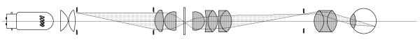
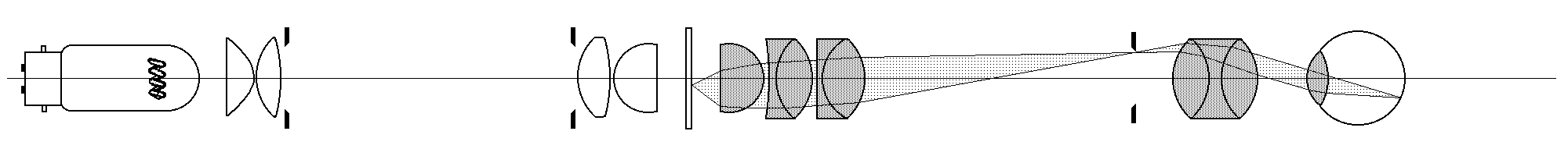
Rather than attempting to illustrate all ray paths using a single optical
diagram, the distribution of light in a Köhler-illuminated microscope is
here represented as four light paths of practical importance to the microscopist:
These diagrams (conforming to the convention that light rays should pass from left to right) extend out of the page to the right for two or three screens (depending upon your screen resolution) enabling a detailed examination of the four ray paths. It is then possible to form a complete impression of what is happening in any section of the microscope by scrolling vertically between the diagrams to compare the ray paths. Images of a given point are formed in the optical train wherever the rays originating from that point cross over. The first image formed will be upside-down; the second will be right way up -- and so on in alternation throughout the system. Path 4 shows that the first (inverted) image of the specimen is formed in the plane of the eyepiece, and the second (erect) on the retina. The retina normally receives inverted images of everyday objects, so the microscope image therefore appears upside down. Adjusting to this in order to follow a moving specimen is probably the first major skill required of the beginner. |
|
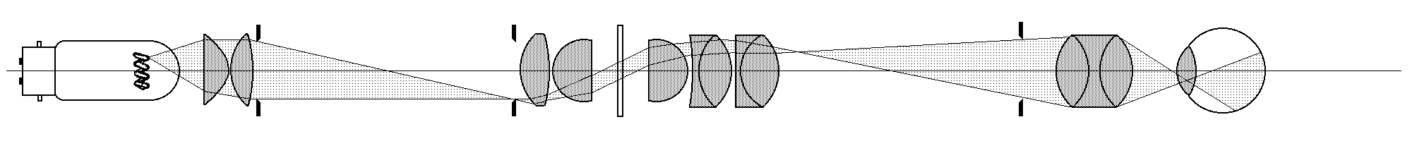
Light from a low-voltage high intensity bulb with a tightly wound grid filament is collected by a large aperture condenser system and focussed to produce an enlarged image of the filament in the plane of the substage condenser diaphragm. This image must be large enough to fill the substage condenser otherwise the back focal plane of the objective will not be filled with light. The diameter of the illuminated field is controlled by means of an iris diaphragm (the lamp field diaphragm) immediately in front of the lamp field lens. |
The condenser accepts light coming from the lamp and focuses it onto the specimen at a far greater angular aperture than the lamp alone could achieve. It is focused to form an image of the lamp diaphragm blades in the plane of the specimen, which means that the microscope field is filled with an image of the lamp condenser lens filled with light. (See next light path). |
S |
This is the most critical component in the system with regard to the quality of the image produced by the instrument. The lens system shown here is a representation of a typical X40 achromat, and in practice has a working distance of about 1 mm. The back focal plane of the objective is shown external to the lens system. Whilst this is true for lower power objectives, the BFP of a real x40 objective is usually within the thickness of the rear elements. |
The second most critical component in the optical system. It further magnifies the image produced by the objective, and on older microscopes, also compensates for any residual chromatic difference of magnification remaining in the objective. The additional optics of the modern infinity-corrected systems are not shown. |
Along with curiosity, the reason for the existence of the microscope. The optics of the eye are simple enough -- it's how the brain makes sense of the weirdly distorted image which falls on the spherically curved surface of the retina and processes it into the familiar images of the external world which is food for speculation. |
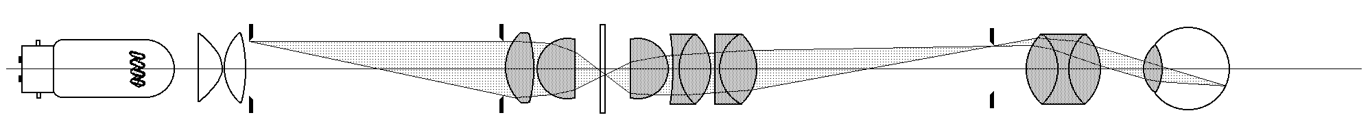 3. Conjugate Images of the Substage Condenser Diaphragm.
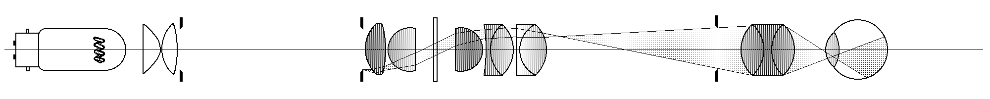 4. Image-forming Rays from the Specimen to the Eye.
|
The term "Köhler-illuminated microscope" describes a system in which all the optical components are design-optimized to work together and are mutually focused upon one another. In this configuration, certain image planes will always coincide. The reader will have noticed that ray path 3 is contained within ray path 1, and similarly, ray path 4 is contained within 2. This demonstrates the fact that in a Köhler setup, the images of the lamp diaphragm and the specimen always coincide, as do the images of the lamp filament and the substage condenser diaphragm. These conditions define the principle and practice of Köhler illumination and give the microscopist control over aperture of illumination (and therefore image resolution, contrast and depth of focus) and over area of field illuminated (giving additional contrast control by reducing glare caused by extraneous light). To the extent that the setup of the microscope departs from the Köhler condition, the above controls are less precise in their effect. If the diaphragms and their images are not correctly located, diaphragm use produces vignetting The next step is the hands-on business of configuring the microscope to the Köhler system.
|