HOME Editorial Articles Specimen Galleries Tutorials Projects Archive Suppliers Links
Worms: Nematodes.
Introduction to Nematode Worms.
Nematode Worm Galleries.
Main
Page

1 of 1
Worms: Introduction Annelids Nematodes Flatworms Parasitic


  The Nematode Worms.

The nematodes are (along with copepod crustaceans) frequently described as "probably the most numerous animals on Earth". Some 80,000 species are described A Typical Nematode 
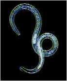
in the literature; possibly a million exist. They live in the soil, in the oceans and fresh water, and are found as internal parasites of most animals and many plants.

It has also been said that if the animals of the world (apart from nematodes) were to dematerialize, their ghostly forms would be recognizable by the populations of nematodes which inhabit their tissues. There are certainly regions of the world where this would be true.

The larger parasitic nematodes are referred to as roundworms, and the smaller parasites as threadworms. They have cylindrical bodies with no trace of segmentation. Chaetae (bristles) are seen only in some of the marine forms. The outer elastic cuticle is shed four times during the life of the worm. The mouth is at or near the anterior end, and the gut is a straight non-muscular tube with an anus at or near the posterior end. A muscular pharynx with a bulbous swelling towards the end is observable in the microscopic forms.

The sexes are separate -- there is no hermaphroditism in the nematodes, but in certain species the females at certain stages reproduce parthenogenetically. They have no blood system, no respiratory system and no cilia. They also have no circular muscle, which means they are not capable of the contractions and constrictions shown by other worms, and typically move in a series of tight S-shaped curves which are fascinating to observe (See movie sequence below).

The parasitic worms in this gallery have been identified to species by scientists at the London School of Hygiene and Tropical Medicine, who also kindly supplied the specimens.

 
  Freshwater Nematodes.

The free-living nematodes are found in moist soil and bodies of fresh water, and are mostly less than a millimetre in length. The population density of nematodes in some marine sediments may reach as much as 20 million per square metre (!) and although these extreme numbers are not encountered in fresh water, almost any sample taken from the debris of the pond bottom will contain numerous specimens.

All microscopic nematodes are remarkably similar in appearance, so they are easily distinguished from other worms, but their identification to species is a task for the expert.
Here is a diagram of a typical nematode (53KB).

Nematode movement.
Click image for larger
movie (84KB).
This movie sequence shows a freshwater nematode worm writhing energetically in a manner entirely characteristic of the microscopic (and macroscopic) nematodes. The constant radius of the body curves can be explained in terms of the tough limitedly-elastic nature of the outer cuticle and the high internal fluid pressure maintained within the body. It is thought that collapse of the digestive canal is prevented by the worm's ability to seal its anus, combined with the pumping action of the pharynx which maintains a countering pressure within the gut. This bodily arrangement is a variant of what biologists describe as a hydrostatic skeleton.
The head end of smaller nematodes is usually blunt, whilst the tail is tapered, and in this case, hooked.

The sequence was originally filmed on 16mm Kodachrome movie film, with synchronized electronic flash as light source. Since the flash duration is 10µsec. the individual frames are without motion blur. The sequence loops (independently of the worm) so that strictly speaking, only the forward part of the sequence shows true worm behaviour. In life, however, the worm can retrace its movements almost exactly as the mirrored half of the sequence.
Darkfield, x200.
Nematode worm in decaying vegetable matter. The head end of a nematode worm found amongst decaying vegetables left for many days in a plastic bag. Dark green liquefied spinach appeared to be the principal component of the sample taken. The worm is surrounded by the remains of disintegrated plant cells, bacteria, and what appear to be fungal hyphae and spores.
(If a passing mycologist could confirm or refute this last observation, Micrographia would be most grateful).
Darkfield, x600.
Nematode worm in decaying vegetable matter. In this sample of pond water, the egg of an unidentified nematode is seen at the centre of the field, surrounded by organic matter containing diatoms and other algal unicells. The developed embryo can be seen coiled within. Hatching will produce a miniature version of the adult worm.
Darkfield, x600.

 
  Brugia.

Brugia malayi (found mainly in the far east) and Wuchereria bancrofti (in tropical and subtropical regions) are the two species of filarial nematodes (long and thin worms) associated with the worm infections known as lymphatic filariasis and its gross manifestation, elephantiasis.

The young infective worms, called third-stage larvae, are transferred to uninfected animals by blood-feeding mosquitoes which have fed upon infected animals. The transfer is not immediate -- the worms which infect the insect take some days to migrate from the insect's flight muscles to the mouthparts (effectively the hollow bottom lip of the mosquito) where introduction to a new host can take place. Here is a diagram of the mosquito's mouthparts.

Once introduced into the host animal, the worms mature into adults of separate sexes in the lymphatic system, usually of the lower limbs. Here the adult worms can cause inflammation and physical obstruction of the lymph vessels. Great swelling of the tissues may result, especially in the legs and the scrotum in men, causing the condition described as elephantiasis.

The large female worms produce thousands of young worms, called microfilariae, that move out of the lymphatics into the blood stream. In the evening these young worms begin to move into the peripheral blood stream where they can be swallowed by mosquitoes biting at night. The microfilariae taken up in the blood meal tear through the mosquito's stomach before migrating to the flight muscles where they settle down to mature into the large infective larvae seen in the photographs below.


Uninfected mosquito feeding.


uninfected mosquito feeding.
The mosquitoes in these pictures are Aedes aegypti, one of the most common vectors of filarial worms used in the laboratory.
The two pictures on the left show uninfected female mosquitoes feeding on human skin. In common with all other mosquitoes, only the females feed on blood -- they need the protein for the production of their eggs.
The top picture shows how the labium, a sheath which normally encases the piercing mouthparts (stylets), peels progressively away from the stylets at this early stage of penetration.

The picture below shows deeper penetration and the way in which the labium remains attatched to the stylets only at the end (the labellum). It is bent in an increasing loop as the stylets penetrate deeper into the skin. When the stylets are withdrawn, the labium will wrap back around them.

The mosquitoes become infected by feeding upon infected animals, and the ingested worms move from the insect's stomach to lodge in its flight muscles. Then, over a period of days, they move from the flight muscles into the space between the walls of the the labium.
The labium is filled with haemolymph (insect blood) and can become filled with infective larvae. When it is bent during feeding, the resulting increase in pressure causes a rupture at the tip (labellum), releasing haemolymph containing worms in a pool around the puncture site. When the mosquito withdraws its stylets, the worms will enter the host bloodstream via the puncture wound.

The pictures in the bottom row are of infected Aedes aegyptii, and they are seen in the process of transferring infective larvae to a new host animal -- in this case, the photographer. (The technique used to obtain the pictures is described in the discussion on photographing feeding mosquitoes in the Projects section).
The first picture shows the tip of a worm emerging from the dislocated labellum. The middle picture shows coiled worms in the pool of haemolymph, and the last shows the enlarged pool around the puncture site just prior to the mosquito's withdrawing its stylets.

All pictures taken with incident multiple electronic flash. x4.
Feeding mosquito with emerging worm. B. pahangi visible at puncture site. Infected mosquito with pooled haemolymph.

An interesting aside in this dismal scenario is that the mosquitoes are also the victims of the worm infection. Those which harbour a particularly large number of microfilariae are usually too weakened and uncoordinated to achieve penetration, thus preventing the development of their eggs.

The photographers who took the pictures of the infected mosquitoes had undergone a course of antifilarial drugs in the week preceding the photo sessions, and in order to reduce even further the risk of contracting an infection, the mosquitoes had been infected with Brugia pahangi, a parasite of cats but not humans. In any case, a successful infection can only result if both male and female worms are introduced, enabling mating to occur in the host bloodstream.

The pictures below are of Brugia pahangi, very similar in appearance to the Brugia species which infect humans.

Brugia pahangi Part of a large tangle of microfilarial worms of the species Brugia pahangi. The picture illustrates the propensity of these creatures to wind around each other, and explains why large numbers of them can so effectively block the lymph vessels they inhabit.
Rheinberg, x100.
Brugia pahangi Part of a single specimen of Brugia pahangi. This tight coiling is characteristic of nematode worms.
Rheinberg, x200.
Brugia pahangi The head end of Brugia pahangi.
Phase contrast, x400.
Brugia pahangi Part of a tangle of Brugia pahangi, showing the heads of two worms.
Phase contrast, x400.

 
  Onchocerca.

River blindness is the common name for Onchocerciasis, an infectious microfilarial disease widespread in Africa and other countries such as Mexico and Guatemala. It is caused by the nematode worm Onchocerca volvulus and is spread by blood-feeding flies of the genus Simulium. Blindness is caused when the worms invade the eyes.

Onchocerca volvulus Part of a tangle of Onchocerca nematodes. The serrations along the length of the body do not indicate segmentation.
Rheinberg, x100.
Onchocerca volvulus Another Onchocerca at higher magnification. Almost the entire worm is filled with developing eggs. Some mature female nematodes are capable of producing 250,000 fertilized eggs each day.
Rheinberg, x200.
Onchocerca volvulus Further magnification on the previous picture. Through the body wall, it is possible to see developing embryos within individual eggs.
Rheinberg, x400.
Onchocerca volvulus Onchocerca -- an egg with a mature embryo.
Phase contrast, x1000.


External Website Here is a link to an interesting article in New Scientist (7 March, 2002) dealing with the commensal bacteria associated with Onchocerca, and the possibility of treating some of the symptoms of the worm infection with antibiotics.


Click to compose email